Breast Thermography (DITI)

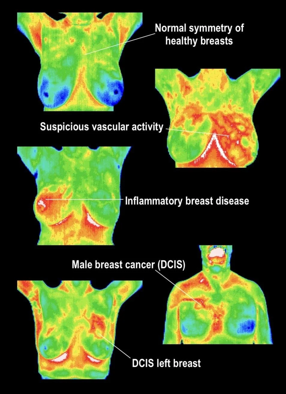
Abnormal Breast Scan

Normal Breast Scan

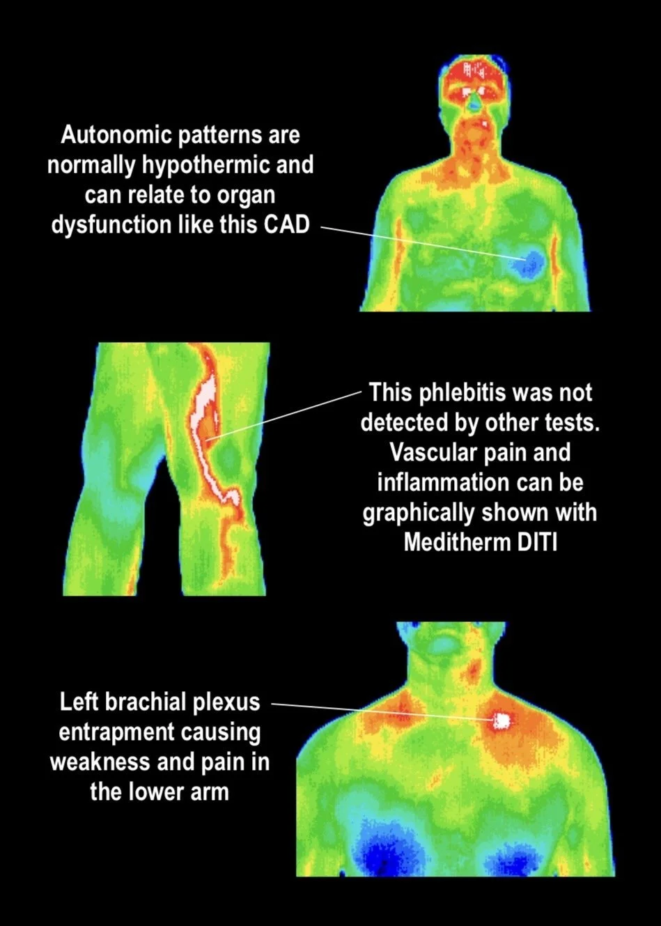
Digital Infrared Thermal Imaging (DITI), commonly known as breast thermography, is a safe, non-invasive imaging technology that measures the natural heat patterns and vascular activity on the surface of the breasts. Using a highly sensitive infrared camera, thermography captures detailed thermal images that may reveal physiological changes associated with inflammation, circulation, and vascular activity.

Unlike mammography, which evaluates the structure of breast tissue, thermography assesses physiological function by identifying changes in heat distribution over time. These thermal images, known as thermograms, can provide additional clinical information that complements traditional breast imaging as part of a comprehensive breast health strategy.

A Comfortable, Radiation-Free Experience

A breast thermography scan is painless, requires no breast compression, and uses absolutely no radiation. The examination typically takes about 15 minutes, making it a convenient option for women seeking a comfortable way to monitor their breast health.

Why Consider Breast Thermography?

Breast thermography can be a valuable adjunctive screening tool by helping identify physiological changes that may warrant additional medical evaluation. It is particularly beneficial for establishing a personal baseline, allowing future scans to be compared over time so subtle changes can be monitored.

Thermography is often chosen by women who want a proactive approach to breast wellness and may be especially helpful for younger women with denser breast tissue, where functional imaging can provide information that complements structural imaging. It is important to note that thermography is not a replacement for mammography or other recommended breast imaging, but rather an additional tool that can contribute to a more comprehensive assessment.

How Thermography Works

Healthy tissues produce predictable heat patterns. Changes in blood flow, vascular activity, and inflammation can alter these patterns and become visible through thermal imaging. An advanced infrared camera detects these subtle temperature variations without touching the body, while specialized software creates detailed color thermograms for clinical interpretation.

Your thermograms are securely stored to establish a personal baseline, making it possible to compare future images and monitor physiological changes over time. When changes are observed, your healthcare provider may recommend additional evaluation using appropriate diagnostic methods.

A Proactive Approach to Breast Health

Breast thermography provides valuable physiological information that can complement mammography, ultrasound, MRI, and clinical breast examinations. When incorporated into a comprehensive breast health program, it offers another perspective to help support informed healthcare decisions and ongoing wellness monitoring.

Breast health matters. Because changes can develop without noticeable symptoms, early monitoring and routine screenings are essential for supporting long-term wellness.